
Growing workload
Cancer incidence and diagnostic complexity keep rising, and every case still needs expert eyes.

AI × MedTech · Singapore
A!Mag builds trusted, explainable AI infrastructure for pathology, so hospitals can detect cancer earlier and pathologists can decide with evidence they can see.
A!Mag MedTechPowered by intelligence. Driven by care.
Scroll to draw
Why we exist
Pathology is the gold standard for diagnosing cancer. Yet hospitals face rising workloads, a global shortage of pathologists, and AI tools that fail to travel between hospitals, scanners and staining protocols. A!Mag builds the infrastructure layer that lets AI earn clinical trust: robust across sites, explainable to clinicians, and validated where it will be used.
Field note
Built to see what the eye can miss.
The clinical problem
Pathology is central to cancer care, but the system is under structural and technical pressure at the same time.
Cancer incidence and diagnostic complexity keep rising, and every case still needs expert eyes.
Experienced pathologists are scarce and unevenly distributed across regions and hospitals.
Most AI models work on the dataset they were trained on, then fail across hospitals, scanners and stains.
Predictions without evidence are hard to trust, hard to regulate and hard to adopt in routine practice.
Existing AI can impress in benchmarks and still fail to deliver trusted deployment in routine clinical practice.
Showcase
Follow a single glass slide through the A!Mag platform, from scanner to signed report.
Scroll to explore
The slide
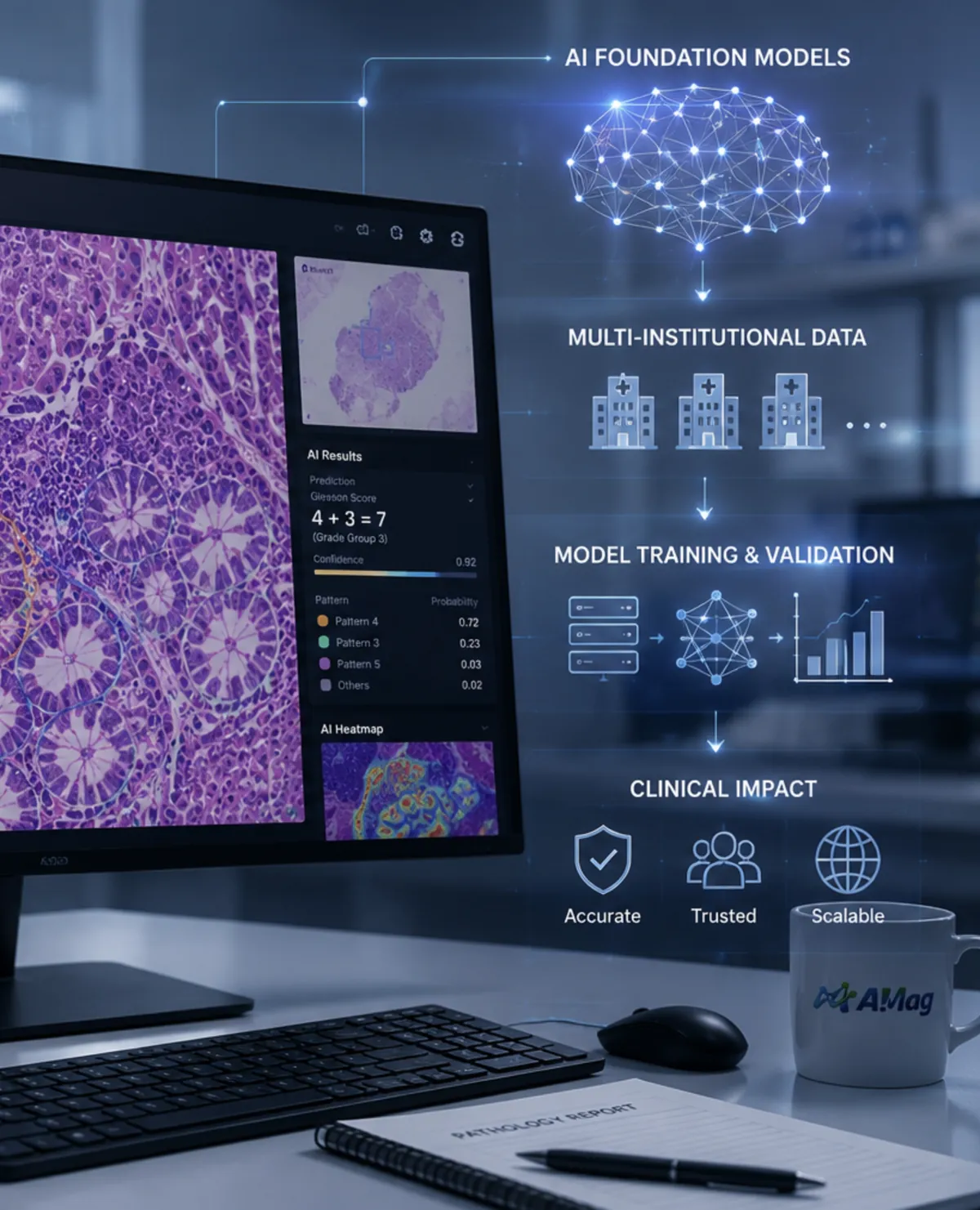
A stained glass slide is scanned into a gigapixel whole-slide image: the raw material of every A!Mag model.
The platform
Models, data pipelines and workflow tools built to generalise across hospitals, scanners, stains and patient populations.

The evidence
Every call comes with evidence a pathologist can see and check: heatmaps and regions, not a black-box score.

The data
Secure, multi-institutional data, so models learn from many hospitals and keep working when they travel.

The pathologist
The pathologist still signs the report. A!Mag magnifies what matters, so decisions are faster and better evidenced.
 A!Prostate
 A!Gastric
 A!Breast
 A!Lymphoma
 A!Colon
 A!Lung
The A! family
A!Prostate leads. Gastric, breast, lymphoma, colorectal and lung follow on the same platform.
Hospitals, partners and investors: the next slide could be yours.
The platform
A full-stack AI pathology architecture, from data to clinical workflow. Each layer is built to generalise across hospitals, scanners, stains and populations.
Whole-slide images, metadata and expert annotations, with quality control at the source.
Self-supervised learning combined with supervised clinical learning and pathologists' domain knowledge.
Evidence regions, similar cases and reasoning support that pathologists can inspect.
Diagnostic modules, quality control and reporting support for specific diseases.
Hospital integration, deployment and monitoring inside existing pathology operations.
Explainable by design
Every A!Mag prediction comes with evidence: the regions that drove it, comparable cases and a reasoning summary a pathologist can check at a glance. Switch on the evidence overlay and change magnification to explore.
Illustration of the interface concept. Not clinical output.
Inside A!Mag
The people, the machine and the platform behind the work. Drag to explore.

Go deeper
The platform, the investment case and the questions we hear most, each on its own page.
The three pillars, the A! product family, the open ecosystem model and how the technology is validated.
Open pageWhy now, the investment highlights, the diversified revenue model and the team structure.
Open pageShort answers for hospitals, partners and investors.
Open pageContact
Hospitals, laboratories, industry partners and investors: tell us what you are working on.
We keep our team private online. Every enquiry is read and answered personally.
We copied it to your clipboard. Send it to the address below, or open your mail app.
Platform & solutions
How A!Mag turns whole-slide images into trusted, explainable decisions, and the products, ecosystem and validation behind it.
What sets the platform apart
A full-stack AI pathology architecture, from data to clinical workflow. Each layer is built to generalise across hospitals, scanners, stains and populations.
What sets the platform apart
Digital pathology image quality control, interactive visualisation and efficiency quantification, with data security and usage authorisation built in.
Consistent accuracy across hospitals and scanners; explainable reports with reasoning and logical analysis; multiscale diagnosis built on pathologists' prior knowledge.
Multiple validated models that answer urgent needs in pathology with reliable AI solutions.
The journey of a slide
Whole-slide images, metadata and expert annotations, with quality control at the source.
Self-supervised learning combined with supervised clinical learning and pathologists' domain knowledge.
Evidence regions, similar cases and reasoning support that pathologists can inspect.
Diagnostic modules, quality control and reporting support for specific diseases.
Hospital integration, deployment and monitoring inside existing pathology operations.
Scroll to move the slide
How it works
An illustrative example of what the pathologist sees: the slide, the model's evidence and the reasons behind a flag. Synthetic image, not patient data.
01
A whole-slide image, viewed at 20×.
02
The regions that drove the call, shown as a heatmap the pathologist can inspect and challenge.
03
A structured draft with the evidence attached. The pathologist decides and signs.
Illustrative · synthetic data
The platform
One platform, every scanner.
Solutions
Open innovation at the core, enterprise value at scale. One architecture supports open adoption and enterprise-grade products for hospitals and industry.
AI infrastructure platform
Foundation models, digital pathology data infrastructure, annotation and knowledge management, quality control, explainable AI frameworks and secure computing environments.
Disease-specific AI products
Clinical applications that fit into routine pathology workflows and are built for regulatory approval, starting with prostate cancer diagnosis and grading.
Services and ecosystem
Deployment, clinical validation, model customisation, regulatory support, education and hospital digital transformation, delivered with clinical and industry partners.
Start with low-friction tools; expand into regulated diagnostic products.
Tap a card to reveal the organ
Digital pathology quality control
Data protection and usage authorisation
Visualisation and annotation platform
Prostate cancer diagnosis and grading
Gastric cancer diagnosis support
Breast cancer pathology
Lymphoma classification and grading
Colorectal classification and grading
Lung pathology
Products are in development and clinical evaluation. Availability depends on regulatory clearance in each market.
Ecosystem
A!Mag uses openness to build ecosystem gravity and captures value through enterprise-grade products. Core infrastructure and selected frameworks will be openly available; validated clinical applications, proprietary models and enterprise services sustain the company.
Working with
Hospitals, research institutes and industry partners build on the platform together. Partner names are shared on enquiry.
Clinical validation
Clinical credibility is central to A!Mag. The technology has been developed in close collaboration with hospitals, pathologists and research institutions in Singapore and China, and evaluated across sites rather than on a single dataset.
Robustness beyond a single site.
Human and AI collaborative diagnosis.
Real-world deployment conditions.
Evidence in the scientific record.
Close connections with major public hospitals and national pathology groups through a national AI digital pathology programme.
Collaborative relationships with leading hospitals and pathology centres, enabling large-scale data access and clinical evaluation.
Hospitals, partners and investors: tell us what you are working on and we will respond personally.
Investors
The trends converging on pathology AI, the investment case, the revenue model and how the team is structured.
At a glance
Why now
The opportunity is to build the operating layer for AI-enabled pathology, beyond any single algorithm.
Hospitals are moving from glass slides to whole-slide imaging.
Representation learning can unlock multi-disease pathology models.
Cancer care needs richer diagnostic and biomarker interpretation.
Rising workload demands scalable decision support.
For investors
A!Mag combines national-scale AI pathology assets, clinical credibility and an open ecosystem strategy into a scalable MedTech AI venture. The company is early-stage and is raising its angel round to anchor value and prepare for Series A.
Founder-led scientific depth, a professional operating lead, and an advisory board of pathologists, AI experts, regulatory advisors and commercial partners.
The deck, data room and team details are shared with qualified investors on request.
Names and CVs are shared on enquiry.
Roadmap
Milestones, not promises: the sequence we are building toward. Dates and figures are shared with qualified investors.
Now
The shared platform foundation, built with hospitals.
Now
Lead clinical product: prostate cancer diagnosis and grading.
Now
Anchoring value with early investors ahead of Series A.
Next
Validation across Singapore hospitals and partner sites in China.
Next
Institutional capital to scale the products and the ecosystem.
Then
Gastric, breast, lymphoma, colorectal and lung on the same platform.
Then
Hospitals, research and industry partners beyond Singapore.
Drag or scroll
Hospitals, partners and investors: tell us what you are working on and we will respond personally.
Common questions
Short answers for hospitals, partners and investors. Anything else, ask us directly.
No. A!Mag is decision support. The pathologist stays responsible for the diagnosis, and every AI output comes with evidence regions, similar cases and a reasoning summary, so it can be checked rather than trusted blindly.
Generalisation across hospitals, scanners, stains and patient populations is the central design goal of the platform, and it is tested through multi-centre and cross-scanner studies rather than on a single dataset.
Not yet. Products are in development and clinical evaluation, and are designed from the start to support regulatory submissions. Availability depends on clearance in each market.
Core infrastructure components and selected development frameworks will be openly available. Validated clinical applications, proprietary models, enterprise deployment, support and regulatory-grade services are commercial.
Data security and usage authorisation are built into the platform through A!Secure, and deployment is designed to fit the hospital’s own environment and governance requirements.
Hospitals and laboratories can join as validation or deployment partners; industry and pharma can partner on research and clinical trials; investors can request the executive deck. Use the enquiry form and we will respond personally.
Hospitals, partners and investors: tell us what you are working on and we will respond personally.